Computer guided implant placement surgery
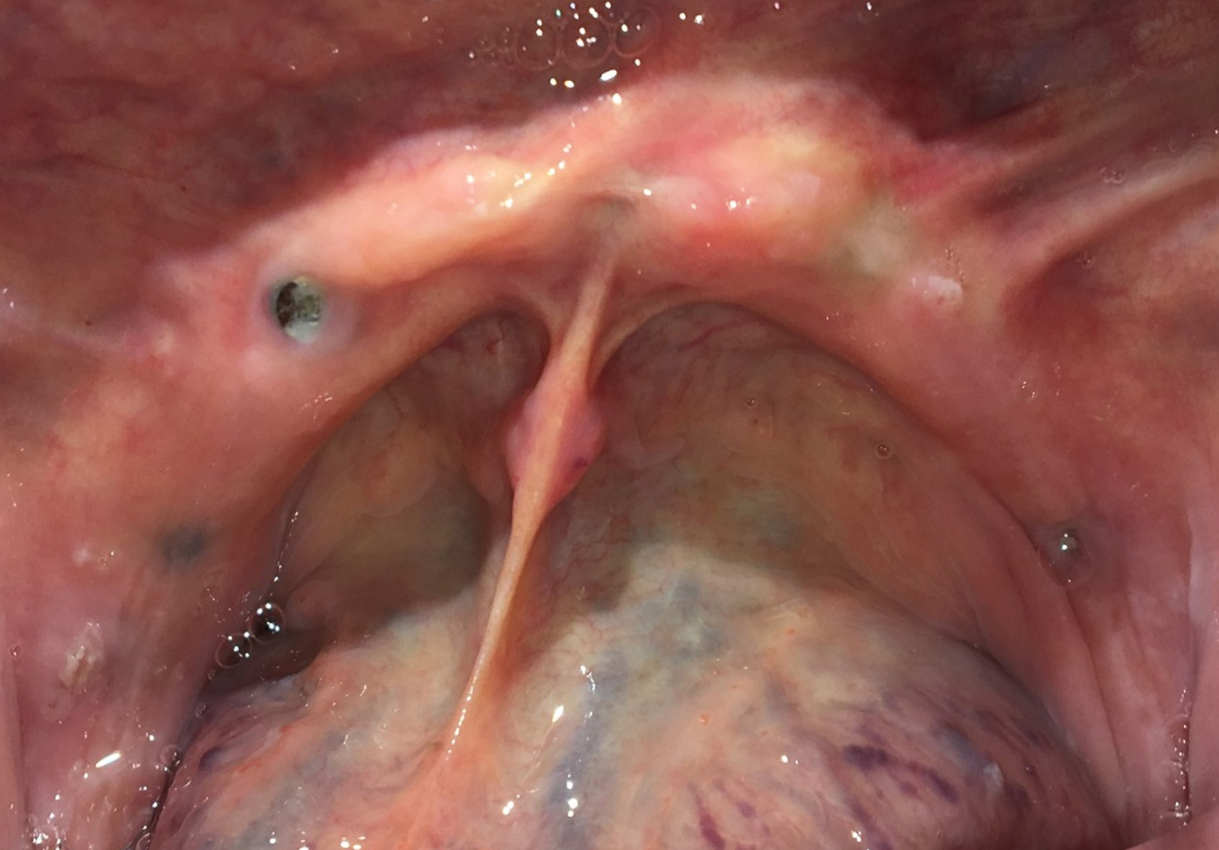
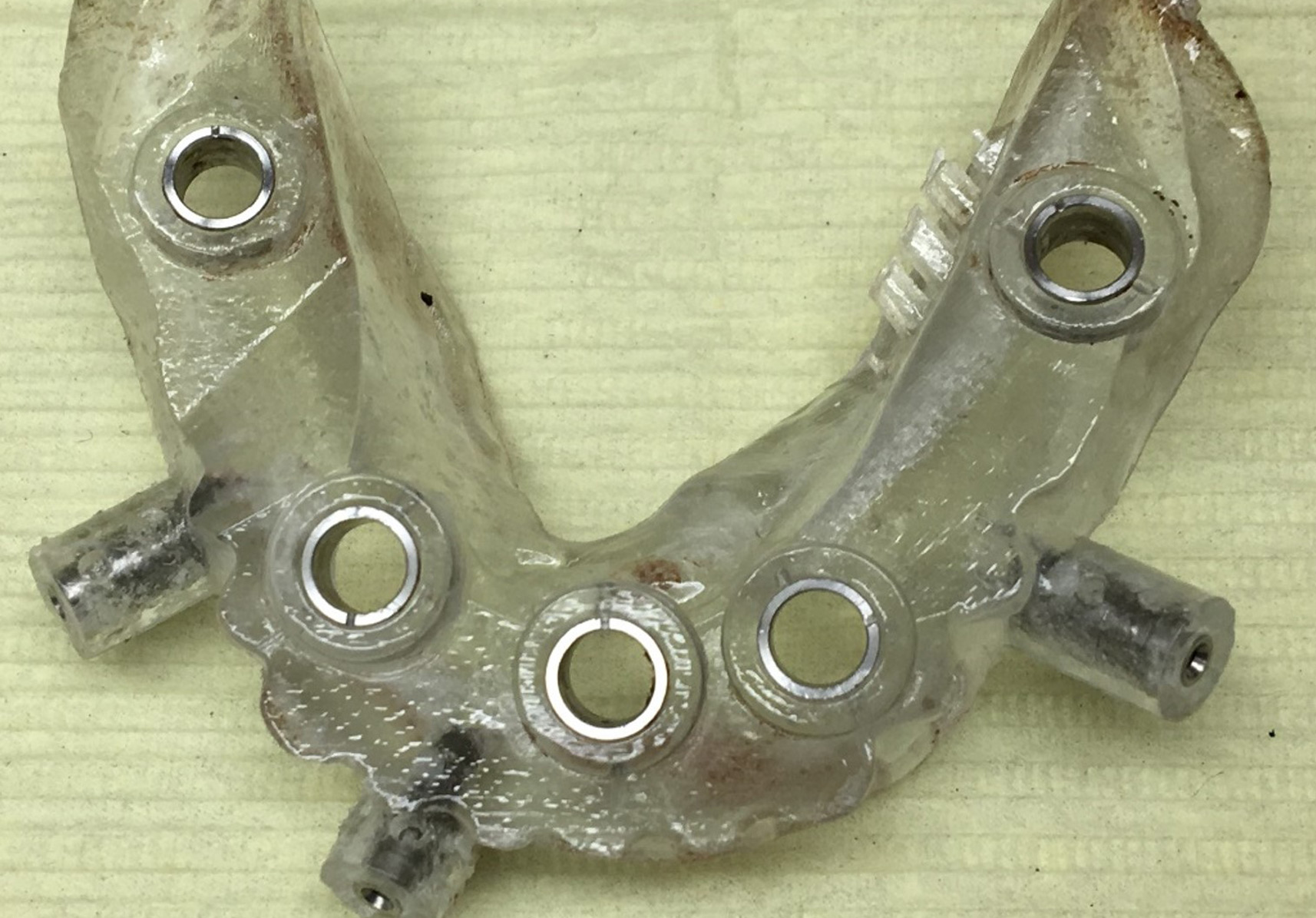
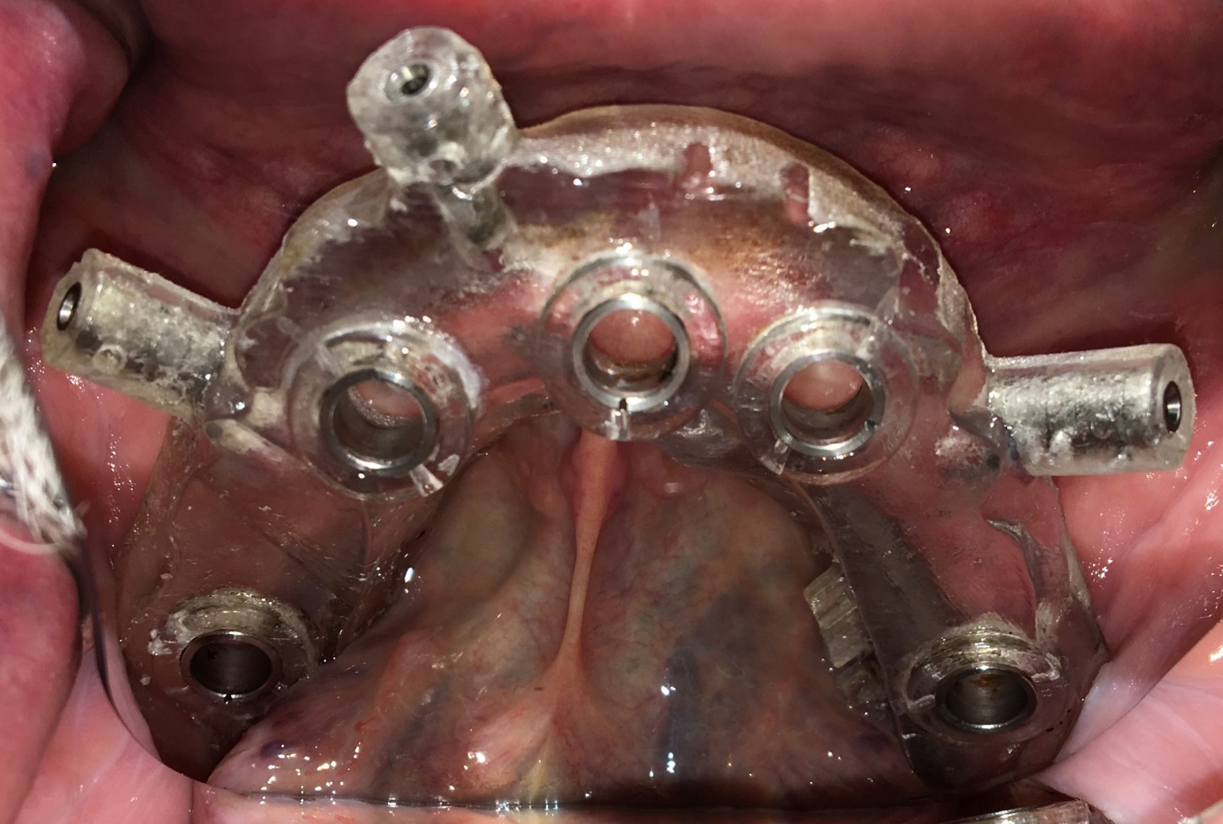
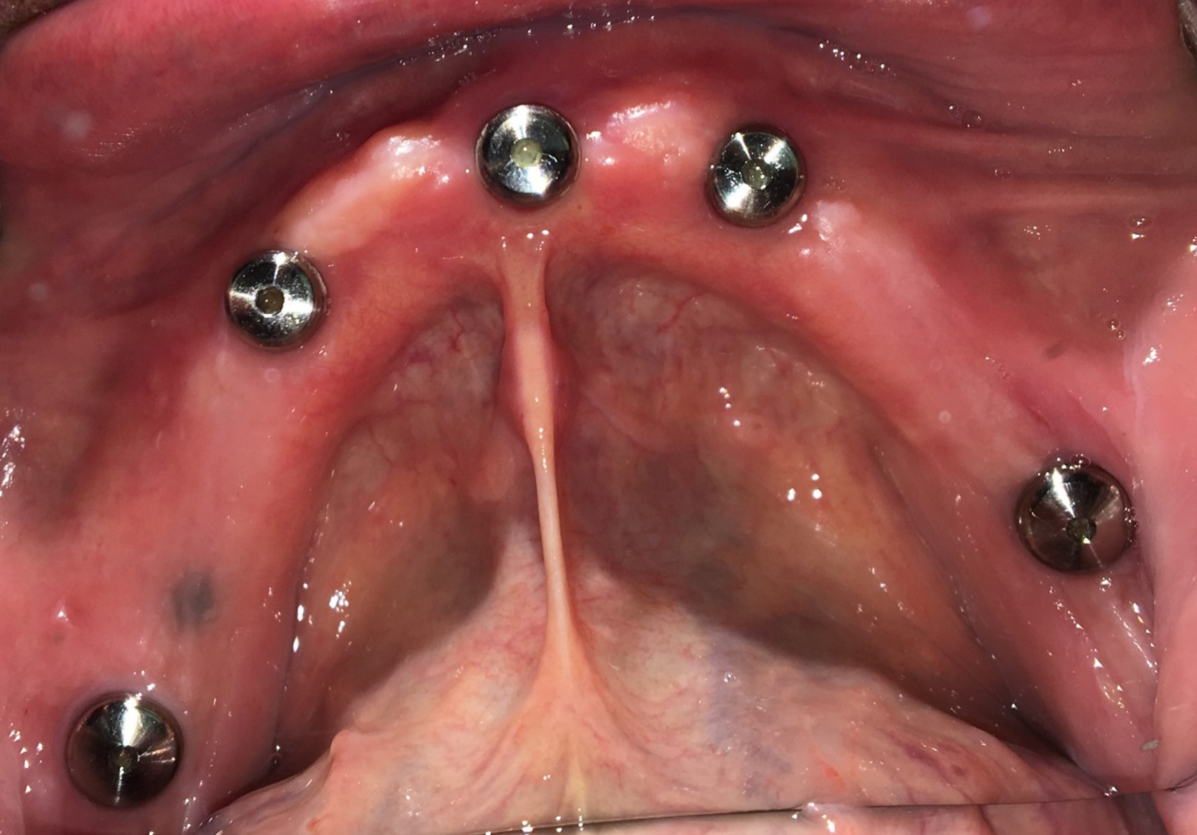
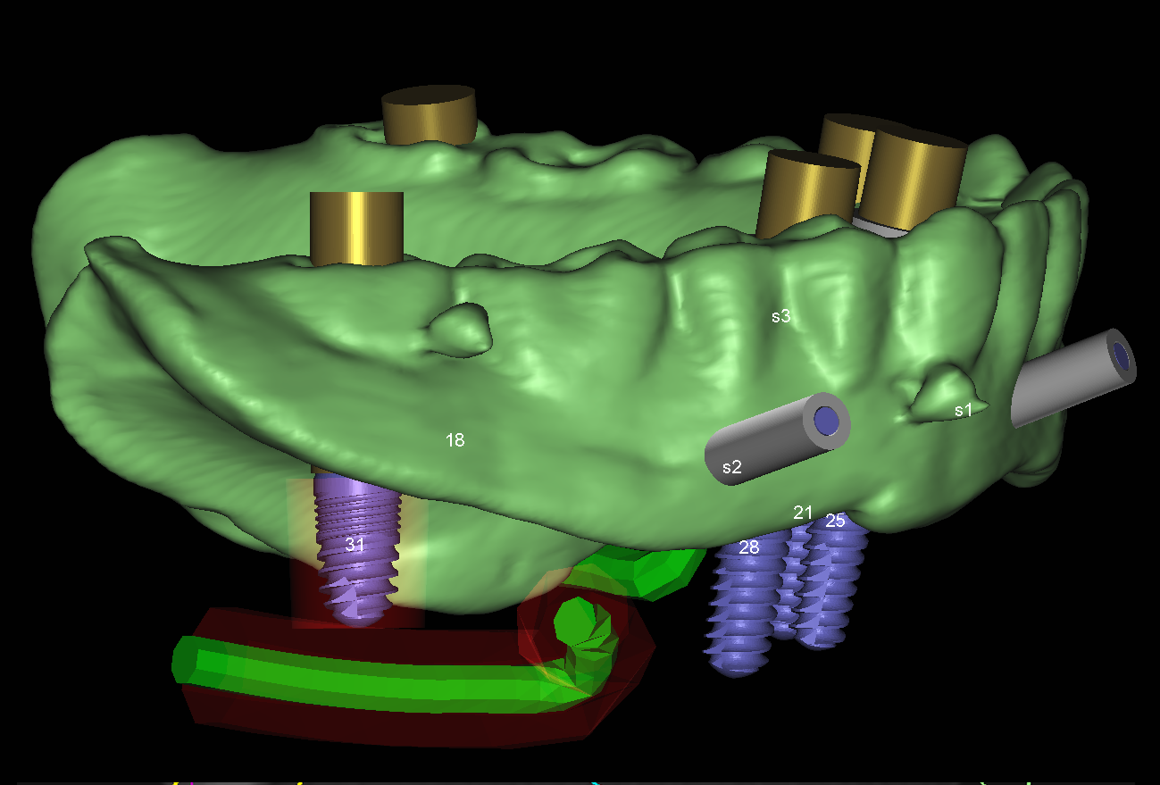
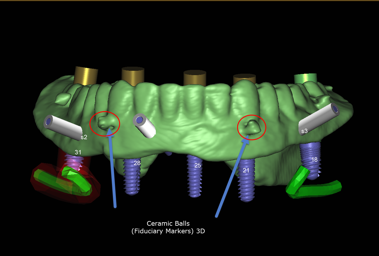
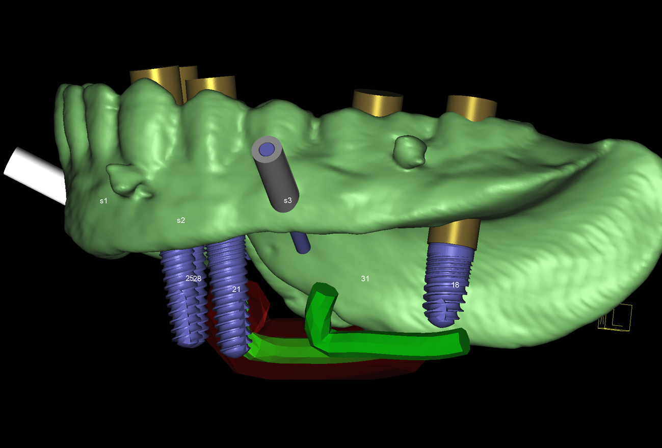
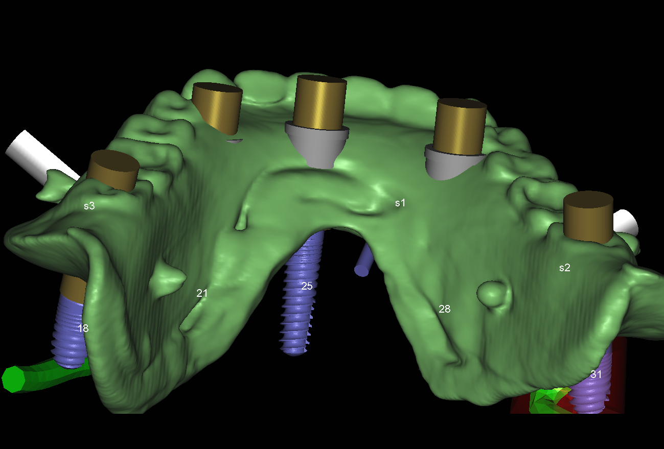
With the advent of 3D imaging technology and surgical armamentaria and technique, our practice provides dental implant placement surgery. Dr. Chipriano uses the principle of computer guided implant surgery, also referred to as digitally navigated implant surgery.
Based mainly on patient demand, Dr. Chipriano invested significantly in nationally and internationally in dental continuing education implantology programs.
Within his dental implants surgery repertoire, Dr. Chipriano has been recognized with several fellowship and mastership awards within implant dentistry. The award criteria are centered on implant specific patient case presentations as well as continuing dental education credit hours specifically designated in the area of implant dentistry.
In 2015, he received fellowships with the International Congress of Oral Implantologists (FICOI) and The International Dental Implant Association (FIDIA). In 2017, he was elevated to mastership the status with the International Dental Implant Association (MIDIA).
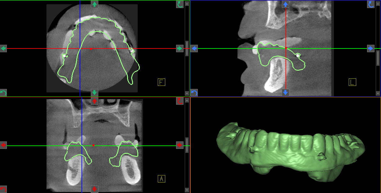
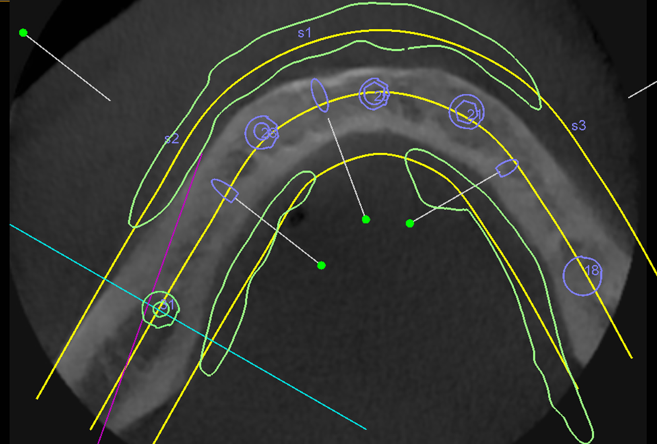
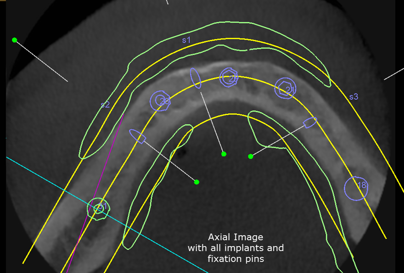
In addition, to develop a precise implant surgical and restorative treatment plan and protocol, our 3D CBCT Imaging Technology offers patients a safe, predictable, and minimally invasive surgery. This is consistently followed by a comfortable and speedy postoperative recovery. To view our 3D CBCT Imaging capabilities, click this link.
Once a treatment plan is devised, a precise surgical guide is fabricated for the implant placement surgery.
Prior to the surgery appointment, the angular path of placement as well as the size(length and diameter) of implants are pre-determined from 3D CBCT scan analysis. This provides for greater predictability in avoiding anatomic structures(sinus cavity and nerves) as well as throughout evaluation of bone availability to allow for implant placement in optimum position and locations.
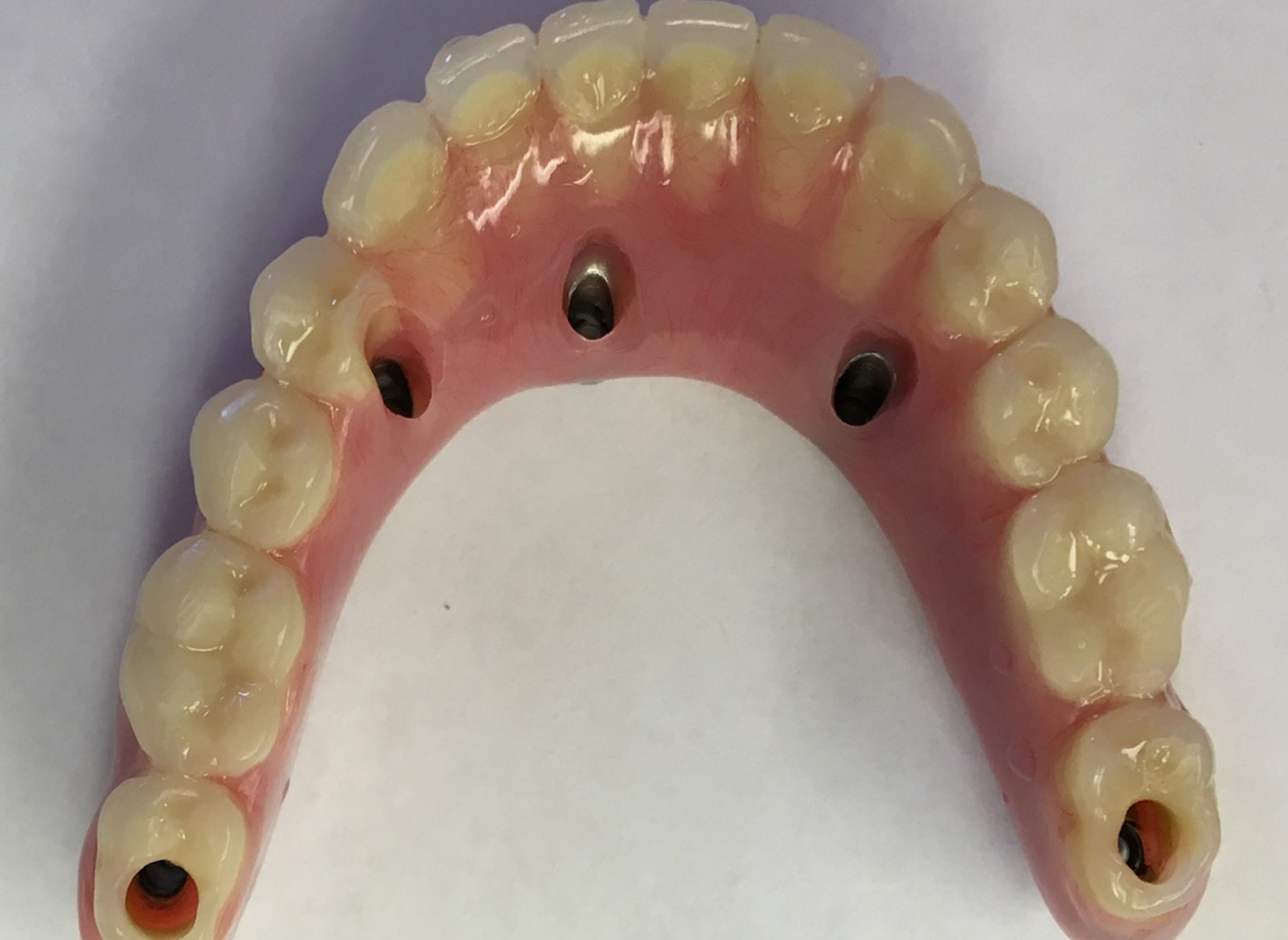
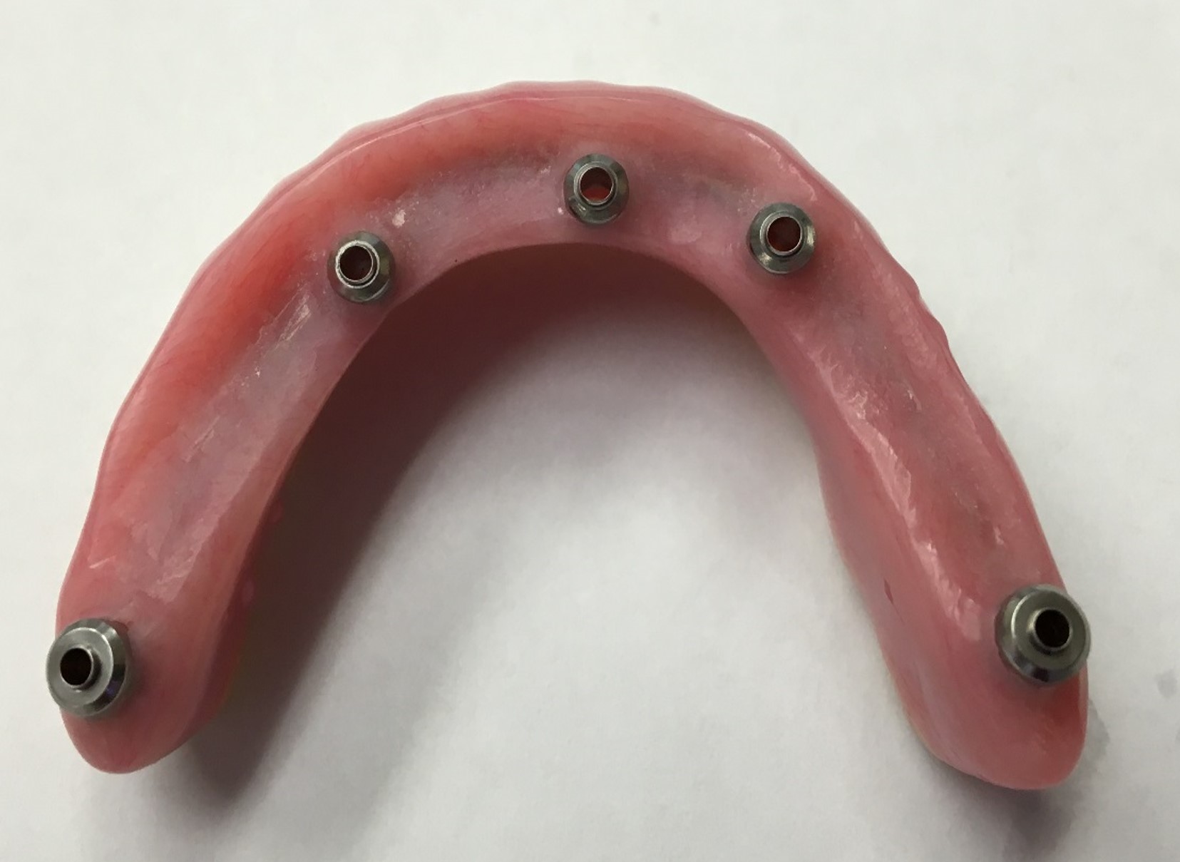
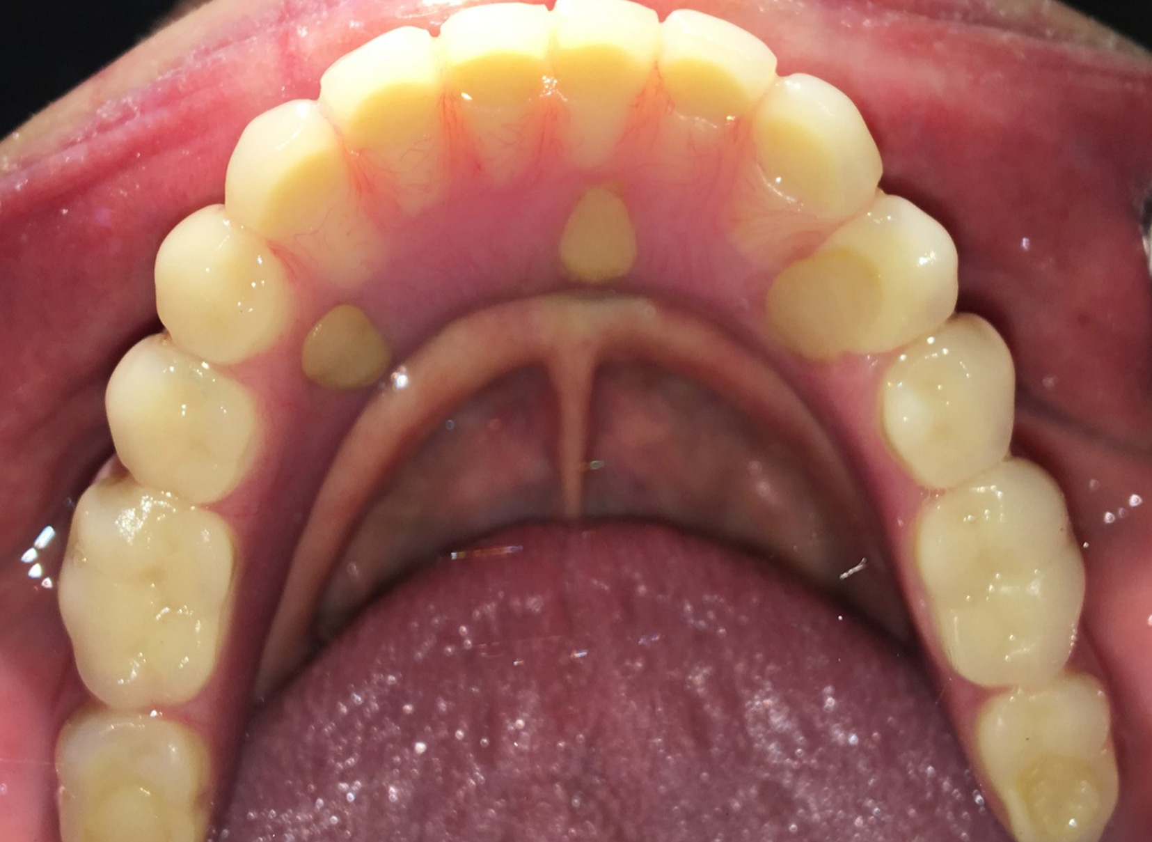
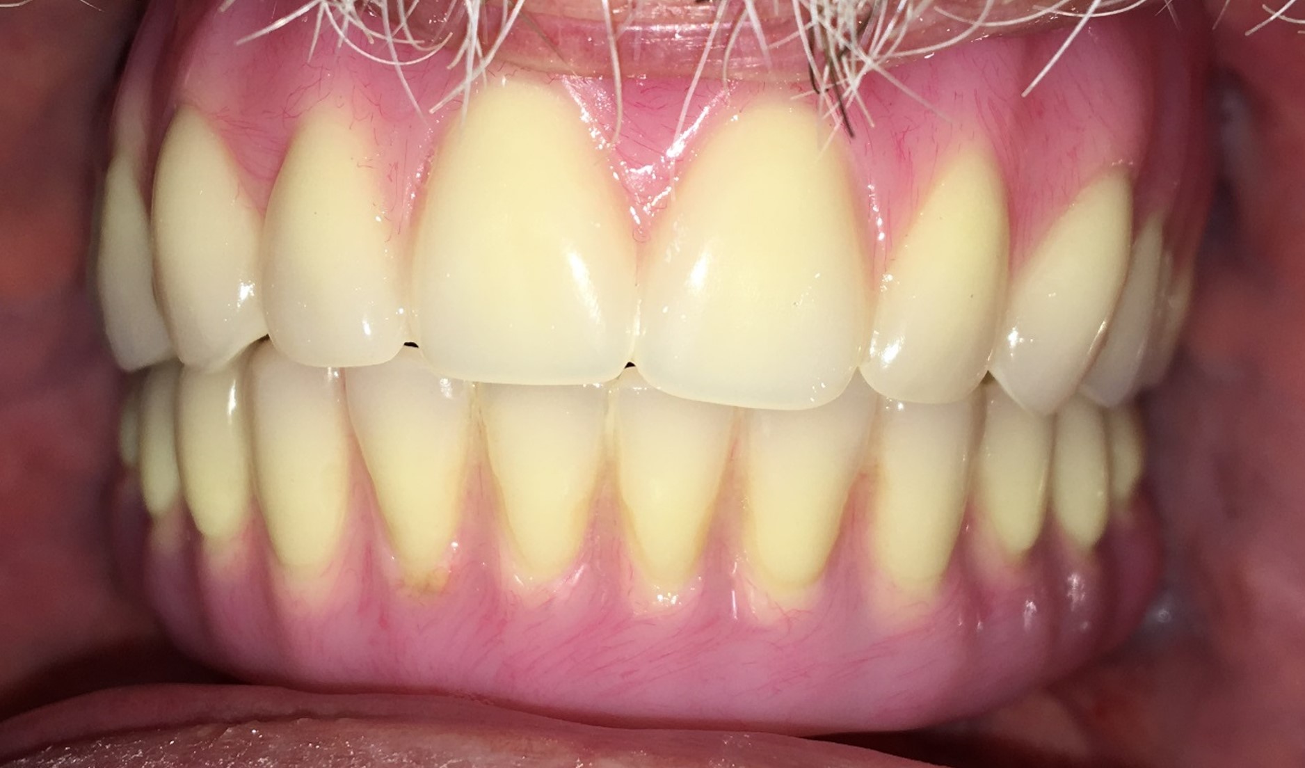
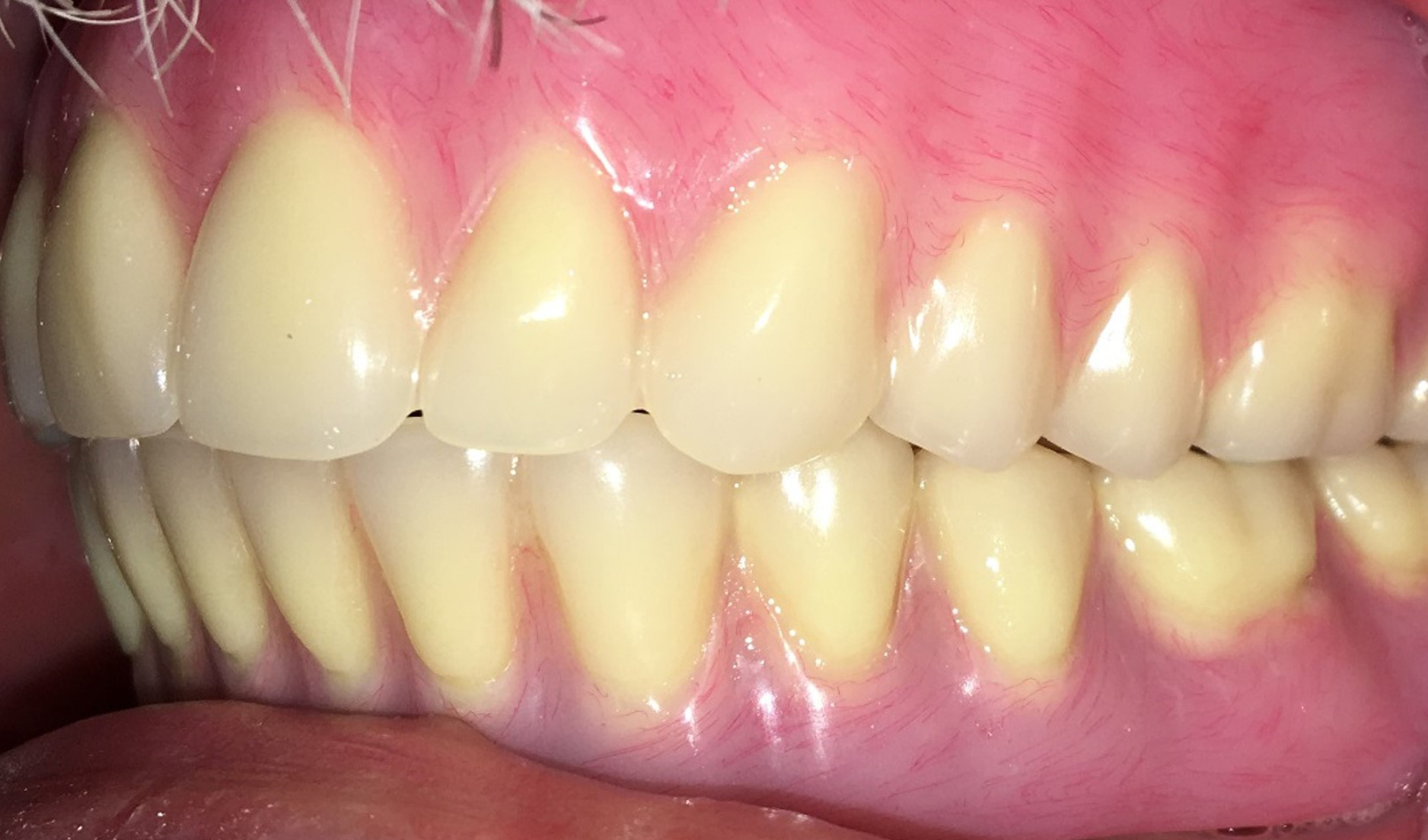
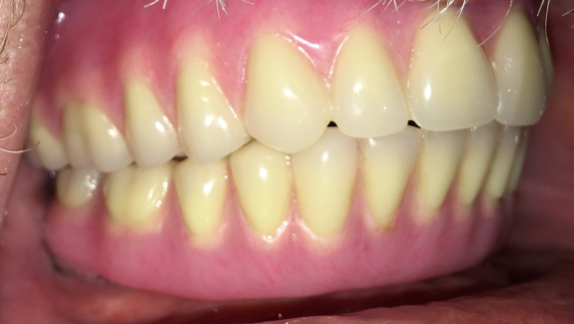
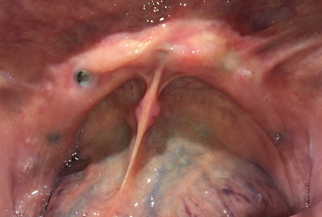
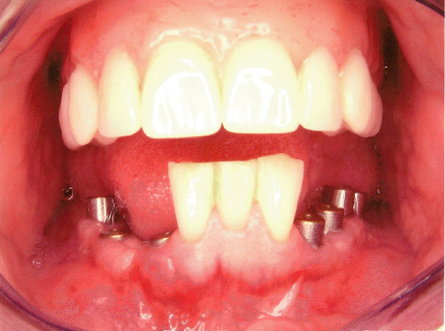
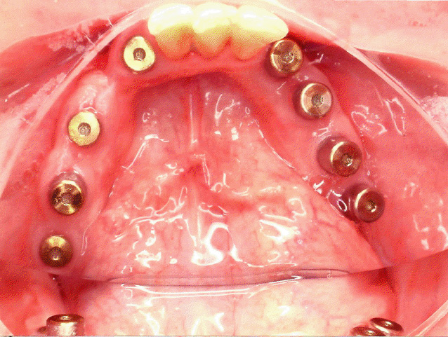
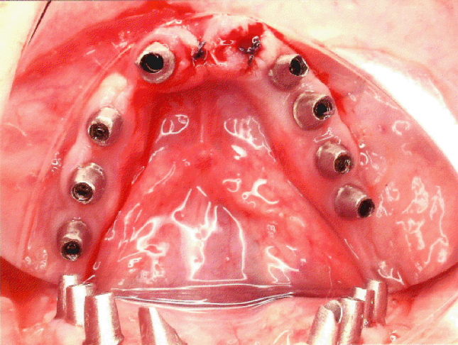
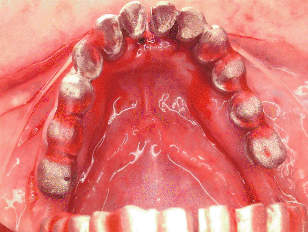
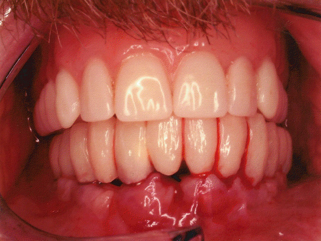
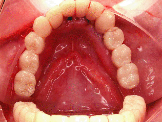
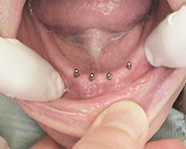
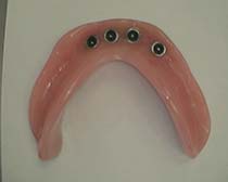
Four months following implant placement surgery, healing copings are placed to allow for a smooth transition from implant site to gum relationship to prosthesis(Crown, bridge, or implant supported denture).
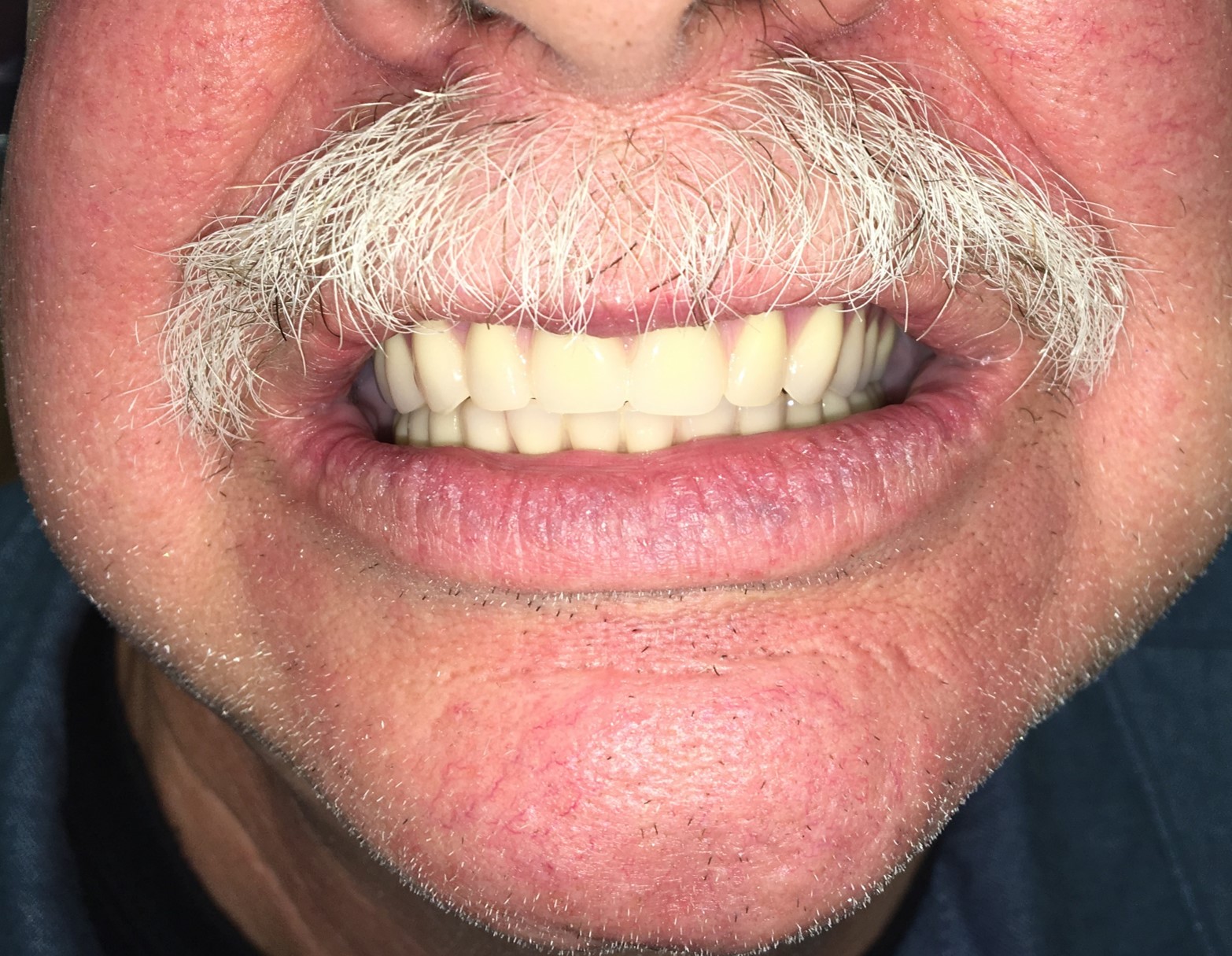
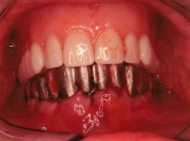
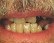
The final prosthesis can then be constructed to allow the patient to regain aesthetics and function.
Because dental implants are often not covered by insurance, we do our best to make this treatment financially possible for our patients. We offer payment by cash, check, or credit card. In addition, third party financing through Care Credit.  Through Care Credit, We offer 12 or 18 months no interest financing options available. These arrangements have been provided on behalf of our patients to prevent the best possible treatment from becoming cost prohibitive.
3D CBCT Imaging
Our practice utilizes state of the art imaging technology to help Dr. Chipriano diagnose potential issues more accurately and provide treatment with unprecedented confidence and precision. This innovative technology is of particular interest in the treatment planning and surgical placement of dental implants.
From the initial 3D image, a thorough treatment plan is established and a surgical guide is fabricated in order to allow for a minimally invasive, conservative, and predictable implant surgery. Click here to view our Computer Guided Implant Placement Surgery.
Our 3D CBCT system allows for precise, crystal clear digital images accompanied by minimal exposure to radiation. We have the ability to control our field of view or scanning area to allow for focusing on broad or specific areas of interest.
Hard Tissue Grafting (Socket or Ridge Preservation)
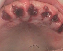
Following the extraction of a tooth, the remaining adjacent bone will shrink in width as well as the depth. Grafting is performed In order to maintain adequate bone levels for the adjacent surrounding teeth as well as to preserve ridge support to better support dentures or a dental implant.
Socket/ridge preservation procedures will allow for bone to be preserved and regenerated in order to facilitate the placement of dental implants. If the patient is remotely interested in restoring a tooth(or teeth) with dental implants, hard tissue grafting is strongly recommended at the time of extraction(s). If bone support is lost with resorption following extractions, a patient may no longer be a candidate for dental implants.
Digital Radiography
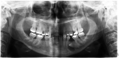
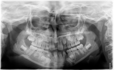
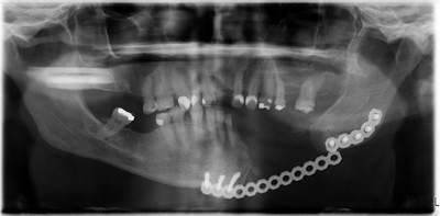
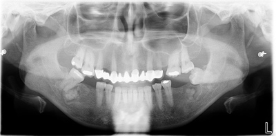
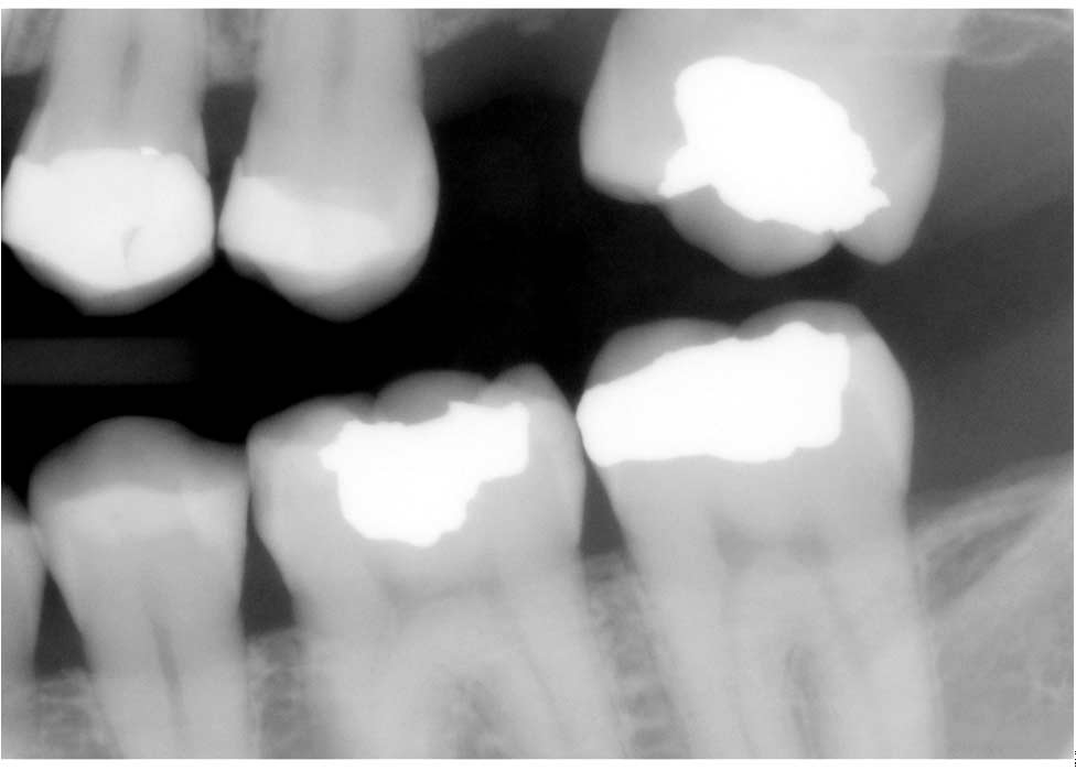
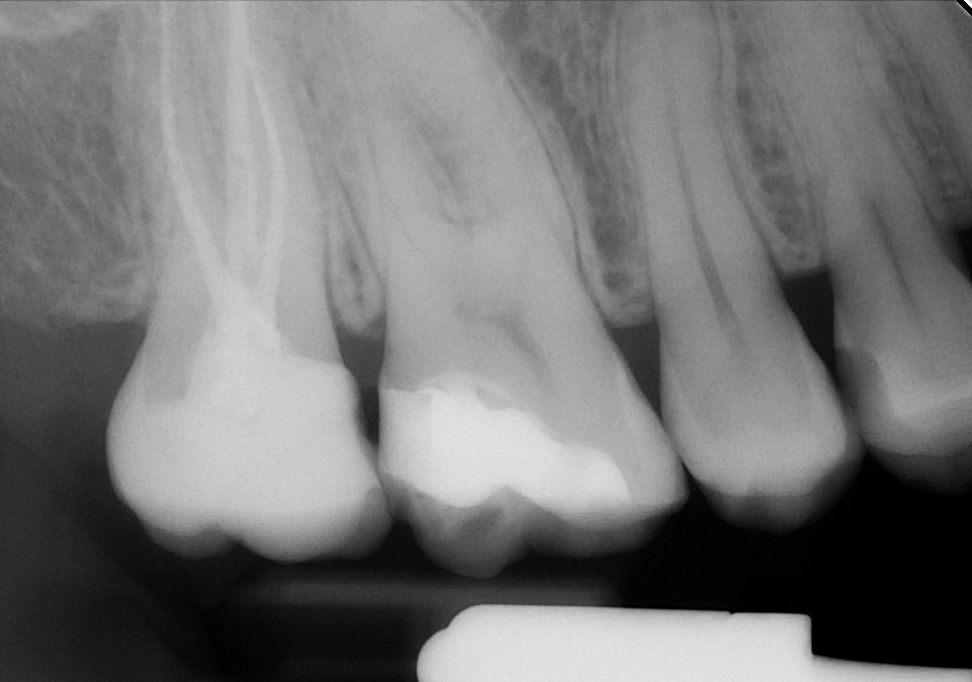
It is not only important to us to provide an exceptional level of care, but we strive to promote patient education and awareness as well. Digital X-rays provide reduced exposure to radiation, less waiting time, shorter appointments, involvement in co-diagnosis, and better understanding of treatment. Digital X-rays are also environmentally friendly. Our office has chair mounted 17" LCD Televisions which allow for images to be clearly displayed and reviewed. The impact is tremendous when a patient has the ability to see what has been occurring dentally as well as what has been done to address it.
Logicon Caries Detection Software
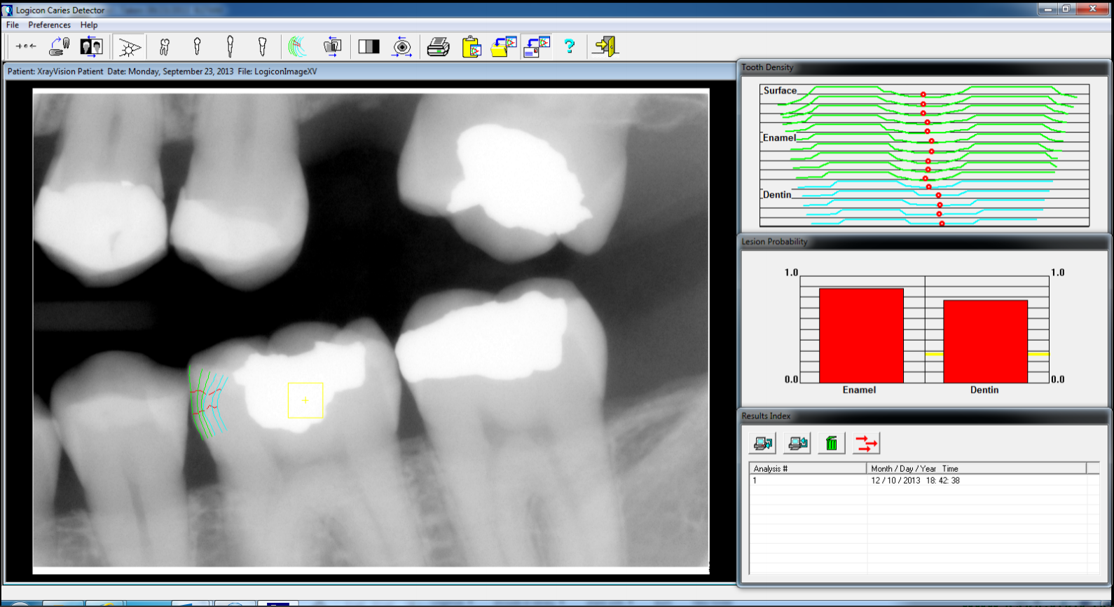
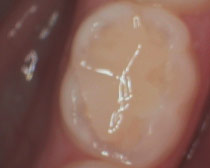
It is our goal to provide conservative and appropriate treatment to all of our patients. In some cases, cavities can be deceiving. We do not want for decay to go unmonitored, undiagnosed, or untreated, so Logicon Caries Detection Software was integrated into our digital x-ray imaging software to provide an electronic second opinion. If decay does not penetrate through the harder enamel surface of a tooth, it is monitored and attempts to remineralize through fluoride is recommended. By using Logicon, we try our best to treat areas of decay which deserve treatment (while they are smaller) and also closely monitor those areas which do not require treatment.
Intraoral Camera
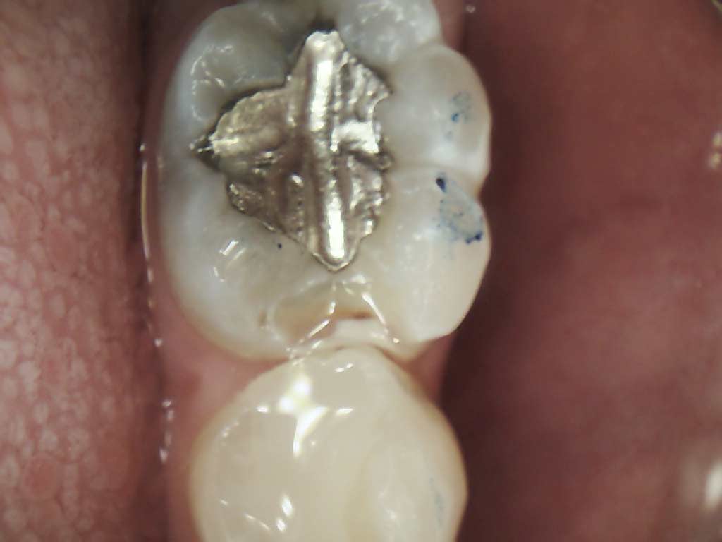
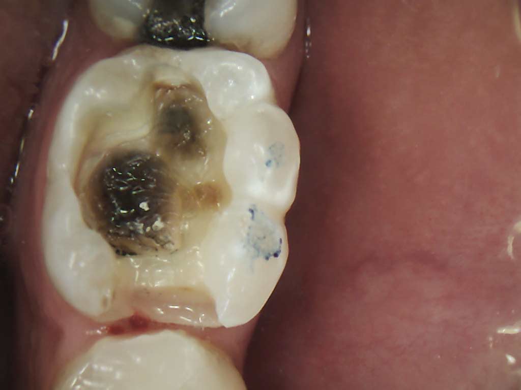
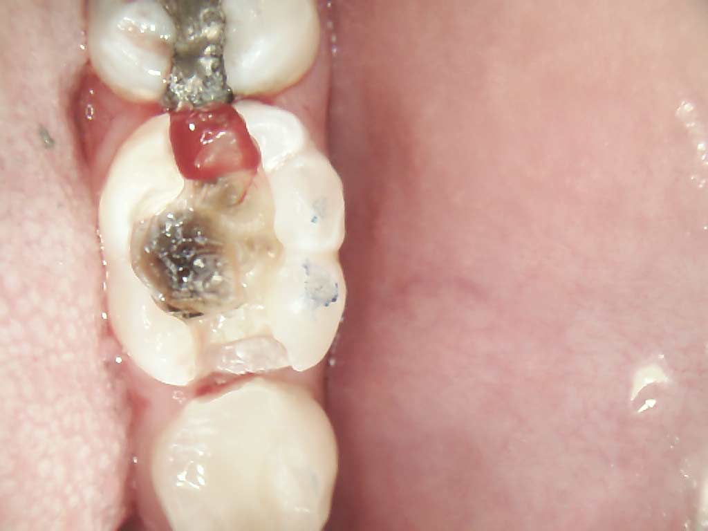
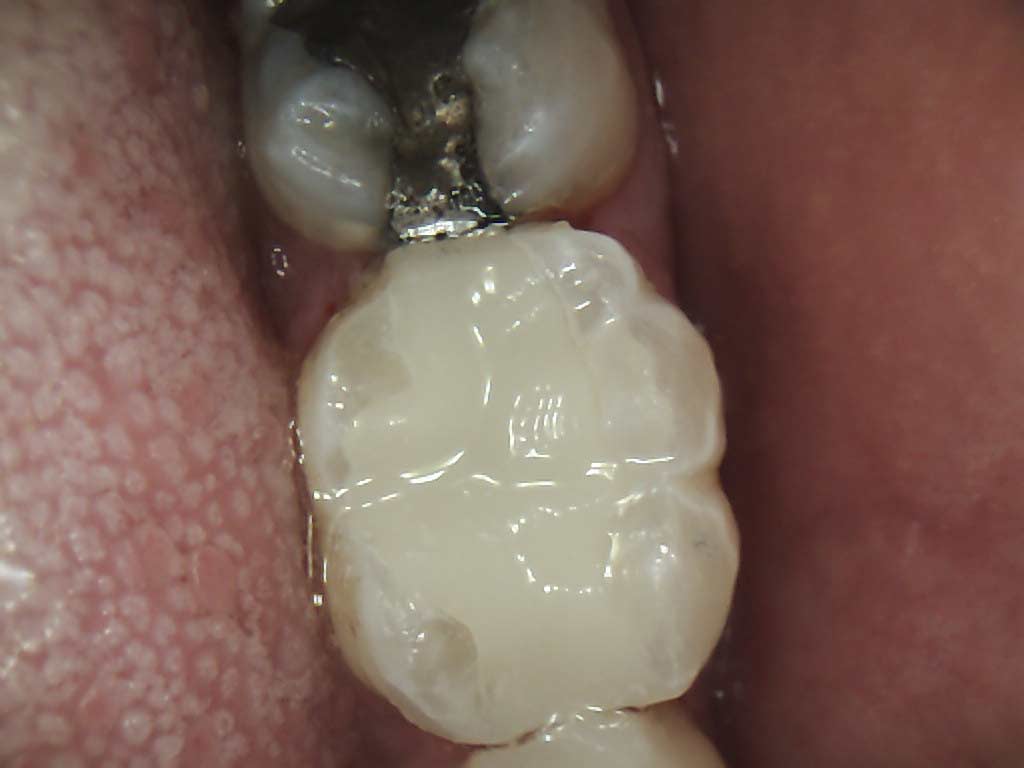
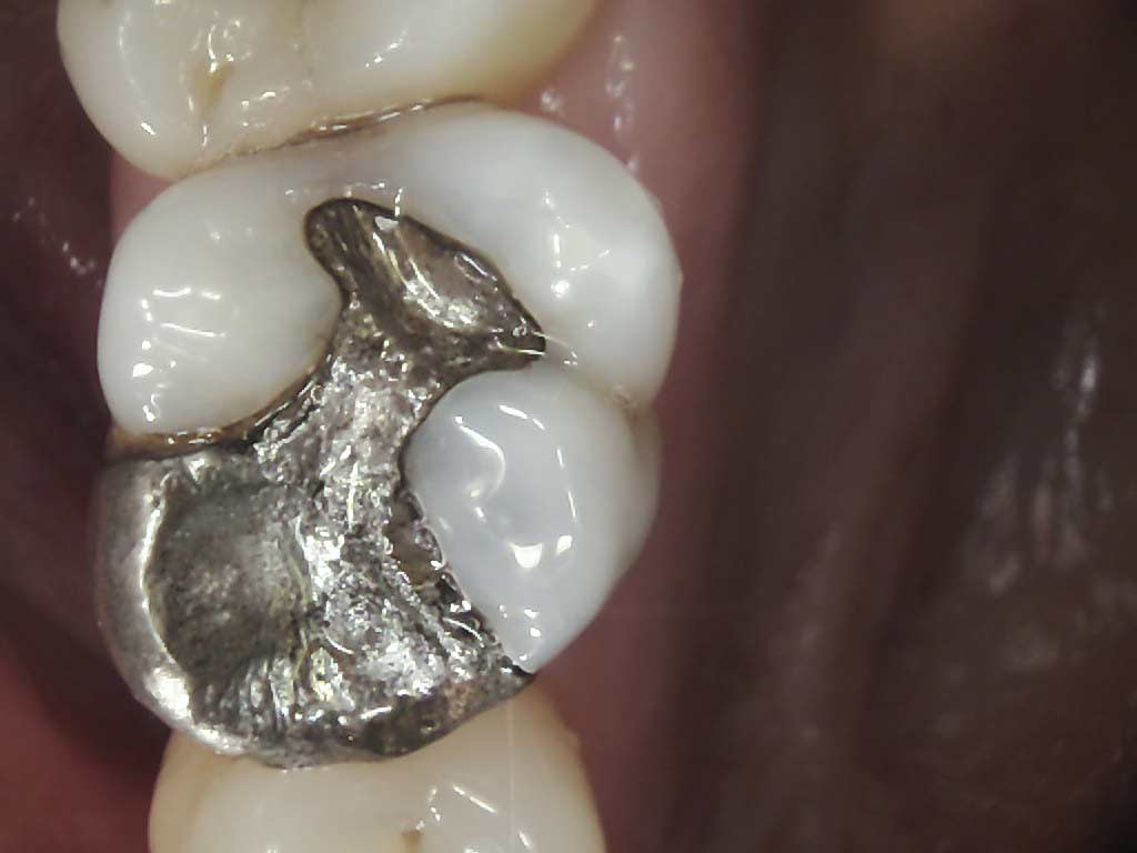
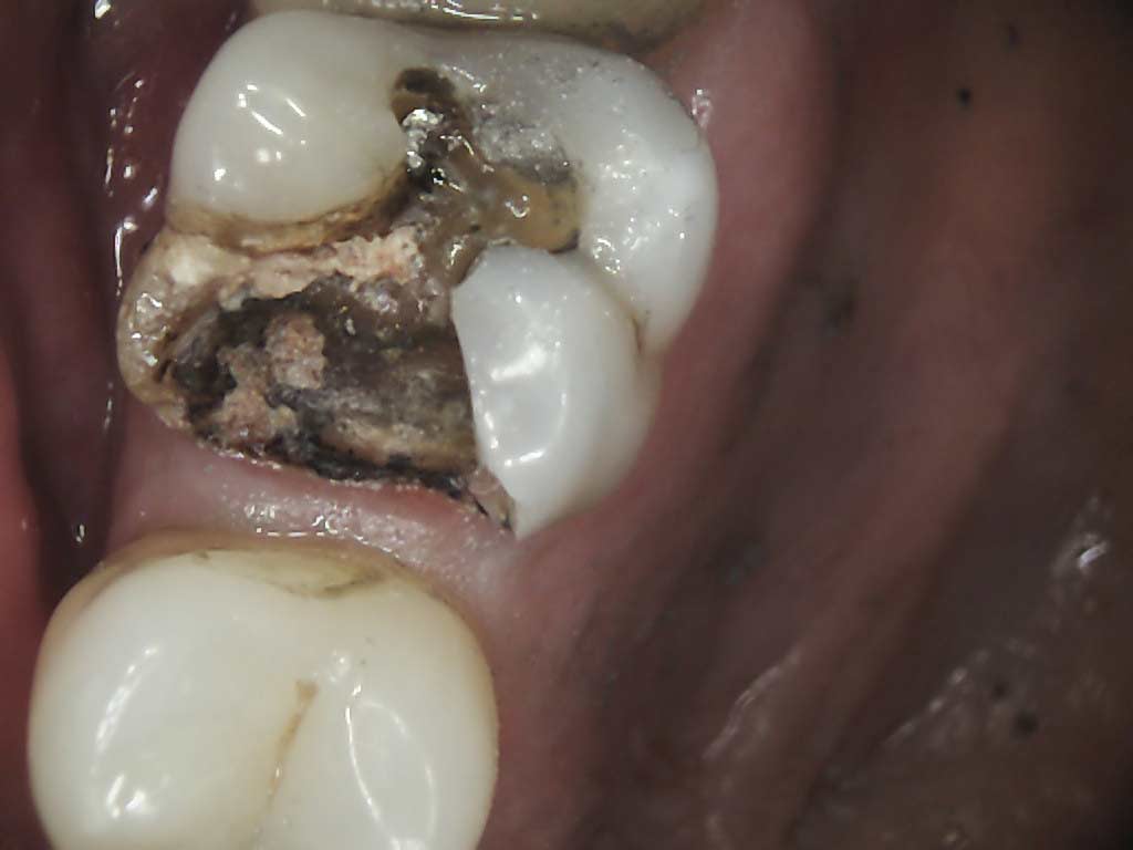
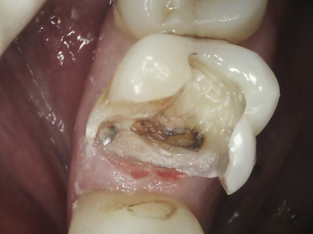
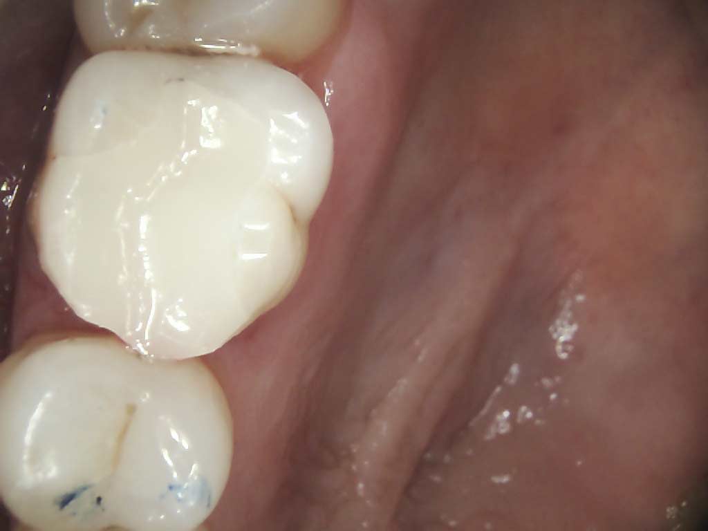
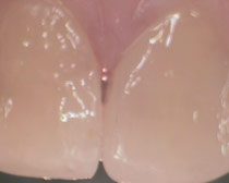
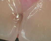
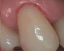
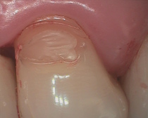
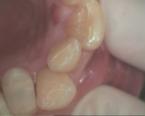
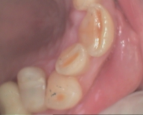
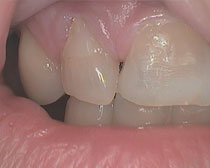
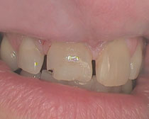
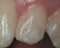
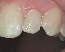
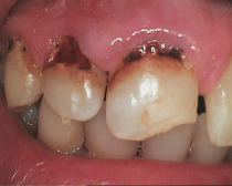
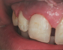
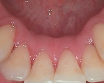
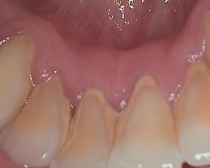
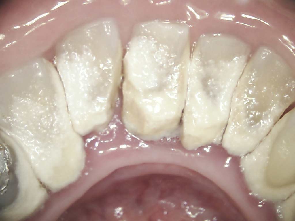
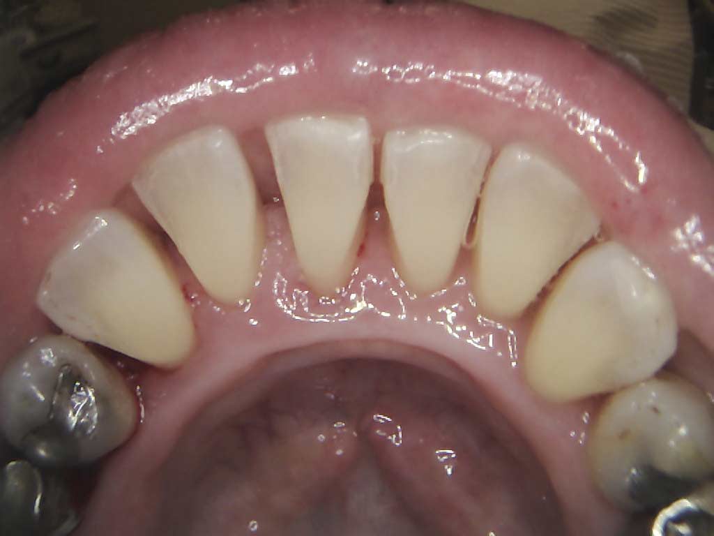
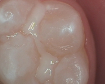
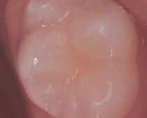
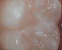
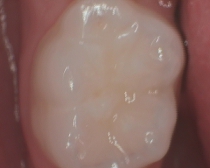
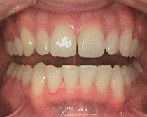
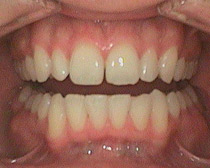
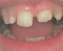
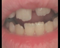
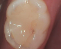
We are pleased to announce that our practice has invested in the state-of-the-art CS 1500 intraoral video camera – the latest word in intraoral imaging technology. This innovative camera is the ideal tool for any oral health practitioner. It offers a superior level of image quality, allowing Dr. Chipriano to capture the perfect shot easily and quickly. The camera allows Dr. Chipriano to easier communicate treatment options to the patients, as well as share images with referring practitioners.
Seeing the clearest possible images helps Dr. Chipriano diagnose and treat patients effectively. Having highly detailed images helps Dr. Chipriano provide quicker and more accurate diagnoses, improved treatment planning and better patient care. The precise level of anatomical detail is essential in determining the best method of treatment.
Benefits include:
•High-resolution images - Dr. Chipriano can view your teeth more clearly
•Wi-Fi connectivity - freedom of movement and better service
•Improved patient care - Dr. Chipriano can perform a wide range of diagnoses, helping you reduce multiple office visits, therefore saving you time and money
We are here to provide you with superb oral care, every time. If you have any questions regarding our technologies, feel free to ask Dr. Chipriano
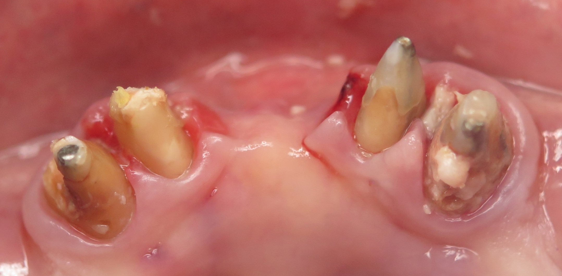
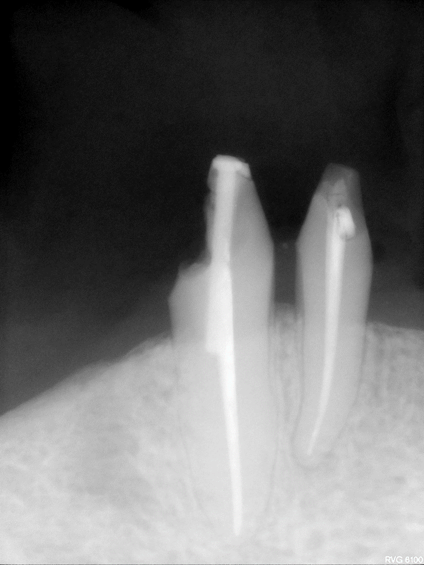
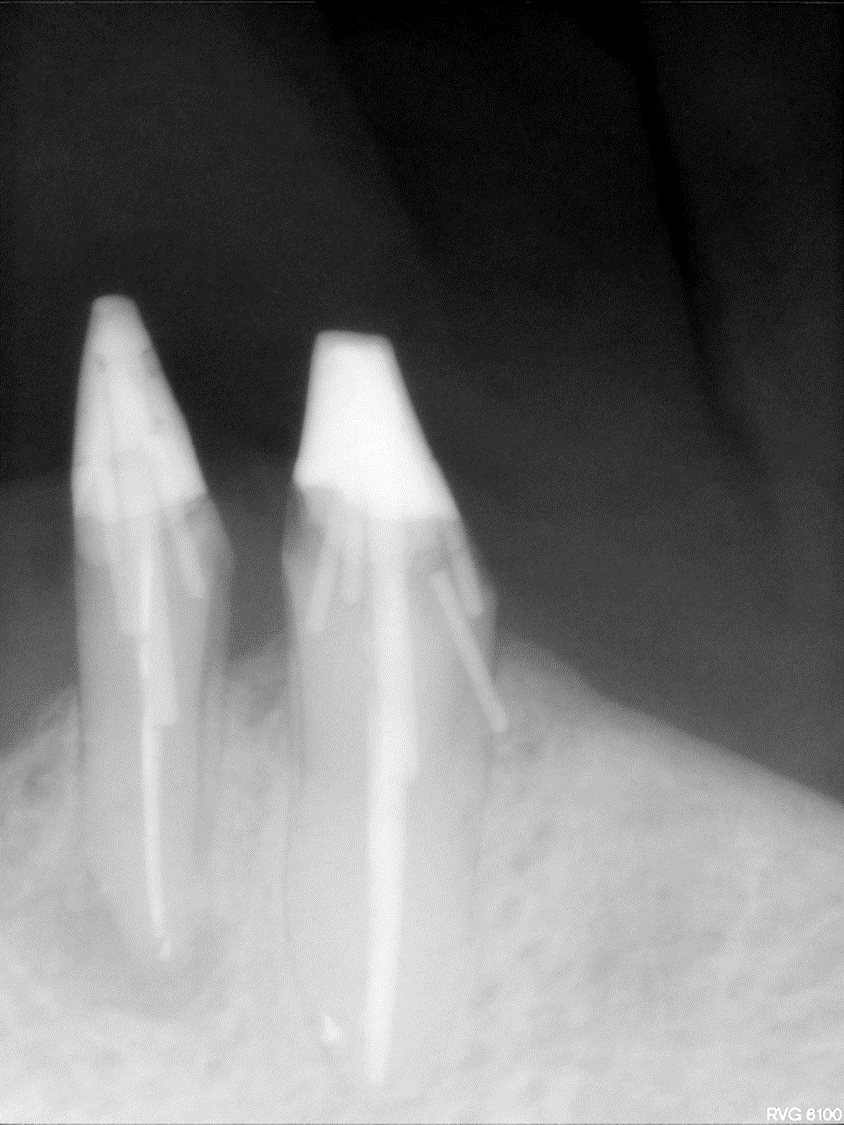
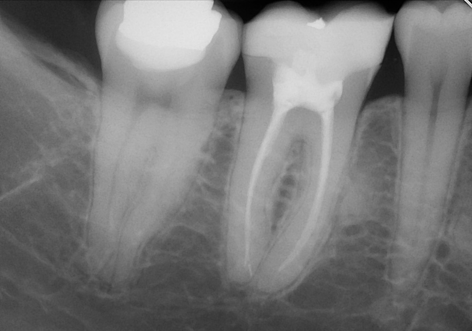
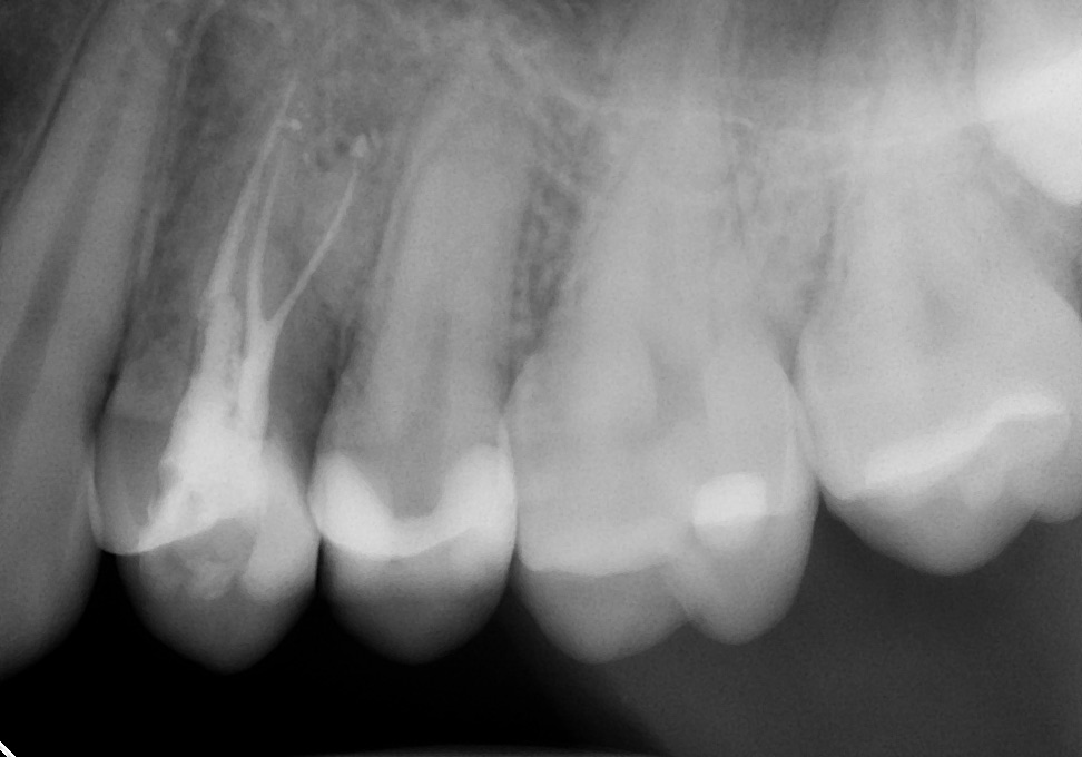
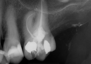 Extremely long, narrow, and curved roots can be cleaned, shaped, and filled with techniques our office utilizes.
Root Canal Treatment
Through the use of rotary endodontics (machine driven instruments to remove problematic nerve tissue from the tooth), computerized measuring devices, and digital x-rays, many root canals can be preformed in one visit, often requiring less than an hour's time. Often in about an hour, a tooth can be treated with root canal therapy and restored with a post and core buildup. Variables include: complexity of the root canal, extent of inflammation or infection, and severity of restorative procedures necessary. By the utilization of warm root canal filling materials, the once problematic nerve canal space is sealed air-tight dramatically reducing chances of re-infection. Root canal treatment by this technique boasts success rates over 90%!


